
Testimonial – Dr. Martin B. Goldstein DMD, FAGD – High Powered Loupes and High Powered Digital Cameras: A Perfect Synergy

High Powered Loupes and High Powered Digital Cameras:
A Perfect Synergy?
Martin B. Goldstein DMD, FAGD
Dr. Martin Goldstein, a fellow of the International Academy of Dento-Facial Esthetics as well as a fellow of the Academy of General Dentistry, practices general dentistry in Wolcott CT, USA. Recognized as one of the Leaders in CE by Dentistry Today for the last 7 years and for his expertise in the field of dental digital photography, he lectures and writes extensively concerning cosmetics and the integration of digital photography into the general practice. He has authored numerous articles for multiple dental periodicals both in the US and abroad. He can be contacted at martyg924@cox.net.
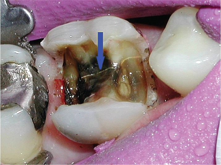
Figure 1 – An easy to spot but threatening midline fracture
Teeth are small. The afflictions that can lead to their demise are even smaller. One such culprit, the intra-coronal fracture, is near impossible to see unless viewed under significant magnification. This is unfortunate as many a root canal treatment might have been avoided had the practitioner been made aware of its presence upon the removal of a failing restoration. While those fractures that have been discolored by overlying alloy or caries are easier to see (Figure 1) others are more subtle and easier to miss; particularly those that reside at the base of a cusp (Figure 2).
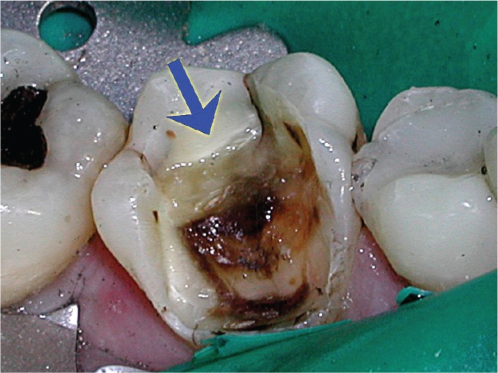
Figure 2 – A more subtle cusp fracture highlighted by a color shift at the base of the cusp
Having begun my own “magnification journey” as many do at the entry-level of 2.0x to 2.5x and having progressed to my current level of 5.5x, I can recall that somewhere in the 4.0x range I began making observations that previously had escaped me. Most notable was a new found ability to actually see the micro-fractures that prior to my enhanced magnification could only be “assumed present.” The flip side of that epiphany was the ability to confidently pronounce a tooth to be “fracture free” within its
coronal structure (above the gum).
Fortunately, my enhanced visual capabilities occurred in parallel with my use of high-resolution digital cameras. Having a hi-res camera chair-side enabled me to do two things. First, after visually detecting an uncovered fracture or suspected fracture site, I could photograph, upload, and enlarge the image on a screen, without fear of pixelization, and thus determine if a true fracture had been detected. Second, I could immediately share my findings with my patient in co-diagnostic fashion. Often this would result in a treatment decision on the fly; specifically, would this be a direct or indirect restoration? Or in laymen’s terms, the “filling vs the cap decision.” As you might surmise, many a planned composite restoration morphed into full coverage restoration upon explanation that the crown solution was more likely to prevent further structural breakdown and thus avert the need for root canal treatment than the less protective filling.
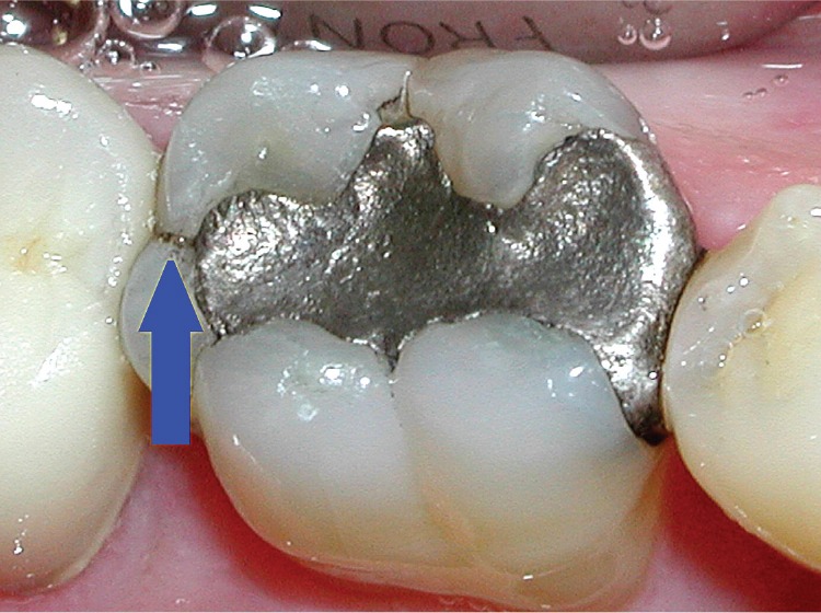
Figure 3 – The ubiquitous marginal ridge fracture
I think that we as dentists and hygienists can all agree that we live in a land of bruxers (tooth grinders) with a baby boomer population replete with amalgam restorations in their mouths. And, if you’ve been practicing long enough, you’ve probably observed that it is extremely common for bruxism-generated fractures to result in both the need for root canal therapy as well as the need for extraction where the fracture rendered a tooth non-restorable. This realization coupled with the ability to see what had been the “unseen,” and photograph it for “all to see,” resulted in my own office becoming much more proactive in our detection, exploration, and restoration of teeth that exhibit clues that trouble is brewing; particularly those teeth exhibiting marginal ridge fractures. (Figure 3) Often an asymptomatic tooth with a marginal ridge fracture will reveal a significant midline fracture upon removal of an older restoration. (Figure 4) Consider removal of such restorations to be “Diagnostic Restoration Removals” or “DDR’s,” allowing the practitioner to intercept fracture-related issues on teeth before they become symptomatic and require endodontic treatment in addition to restorative work. More often than not, all parties will benefit.
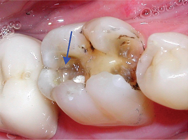
Figure 4 – A more threatening fracture revealed that timely restoration can solve
-Martin B. Goldstein DMD, FAGD
Note: photos pulled from: What Your Eyes Can’t See…A Patient’s Guide to Hidden Tooth Decay and Micro-fractures by Dr. Martin Goldstein, available from Photomed International, www.photomed.net